

Investigation
Sonography
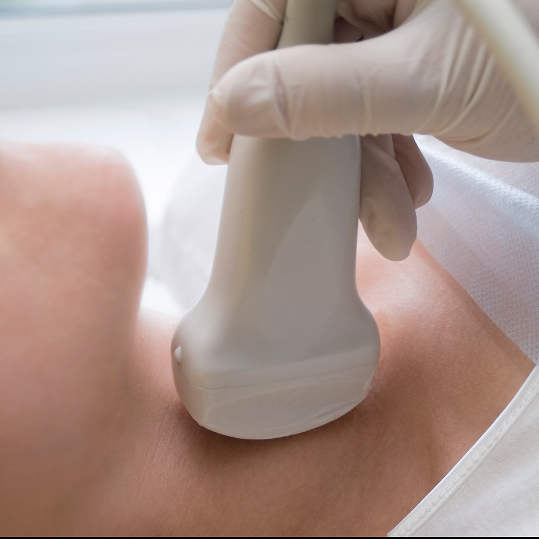
Diagnostic sonography (ultrasonography) is an ultrasound-based diagnostic imaging technique used for visualizing subcutaneous body structures including tendons, muscles, joints, vessels and internal organs for possible pathology or lesions.
Abdominal sonography detects pathological changes in 8 main areas: liver, intrahepatic duct, common bile duct, gallbladder, kidney, hepatic portal vein, pancreas and spleen.
Pelvic ultrasound detects bladder, ovaries, uterus, cervix and fallopian tubes of a woman. It also covers the bladder, prostate gland and seminal vesicles of a man. Pelvic ultrasound can be done in three ways: transabdominal, transrectal, and transvaginal.
Breast sonography is an effective diagnostic tool for cysts or benign fibromas in breast. It is mostly used for young females, females before the menopausal age and females with high density mammary glands.
Prostate sonography is an early diagnostic tool for prostatitis or suspicious prostatic tumour in men. It has a higher sensitivity, consistency and accuracy of diagnosis.
- Detection of fatty liver. When the fat content, particularly neutral fat in the liver increases excessively, liver functions will be impaired. This is called fatty liver.
- Detection of liver cysts. It is usually caused by congenital abnormalities of the biliary tract.
- Detection of renal cysts. If the kidney contains too many cysts, kidney functions will be impaired. Severe polycystic kidney may develop renal failure.
- Detection of liver haemangioma, biliary stones or sediments and gall bladder polyps.
- Effective tool in diagnosing cysts or benign fibromas in women.
- Effective tool in diagnosing prostatic tumor, prostatitis (prostate inflammation) and vesical calculus in men.